Hip Joint Muscles Diagram : Hip - Joints and More - Flexion of hip and vertebral column.. The hip muscle diagram below shows a number of. Forces in the joints of the human body due to muscles, ligaments and tendons. In human anatomy, the muscles of the hip joint are those muscles that cause movement in the hip. Laterally rotates the the thigh at the hip joint. Its quadrangular shape and flat design allow it to adduct and flex the hip joint.
The movements that can be carried out at the hip joint are listed below, along with the principle muscles responsible for each action Hip joint is ball and socket joint that connects axial skeleton with lower limb. Human anatomy diagrams show internal organs, cells, systems, conditions, symptoms and sickness information and/or tips for healthy living. Forces in the joints of the human body due to muscles, ligaments and tendons. Iliopsoas, tensor fasciae latae, sartorius, and rectus femoris muscles.
Hip Surgery Northampton - Hip Conditions Treatment Midlands from www.hip-and-knee-surgeon.co.uk Prime movers cross hip joint anteriorly: The hip joint is located between the head of the femur and the acetabulum of the pelvis on each side. Psoas major, iliacus and rectus femoris learn more about the hip joint by exploring our articles, video tutorials, quizzes and labelled. Related online courses on physioplus. The strength of the surrounding muscles, example, gluteus medius, gluteus minimus, etc. The following diagram illustrates the actions of the terms adduction, abduction, flexion and extension at the different joints. Human muscle system, the muscles of the human body that work the skeletal system, that are under voluntary control, and that. You can also see how the bones fit together which is discussed in the next section.
Globular end of the femoral neck.
Human muscle system, the muscles of the human body that work the skeletal system, that are under voluntary control, and that. Adductor longus, inguinal ligament, sartorius. Microscopic anatomy of skeletal muscle. Flexion of hip and vertebral column. It bears our body weight while we sit, stand, walk, or run. Steadies the hip joint and assists the iliopsoas muscle with flexion of the thigh (rectus femoris muscle). Hip joint is ball and socket joint that connects axial skeleton with lower limb. Prime movers cross hip joint anteriorly: The hip joint is located between the head of the femur and the acetabulum of the pelvis on each side. Knee assessment and hip mechanics learn how hip and pelvis mechanics can influence the knee powered by physiopedia start course. The following diagram illustrates the actions of the terms adduction, abduction, flexion and extension at the different joints. Want to learn more about it? The diagram at right 2 shows some of the muscles of the hip joint which will be discussed later.
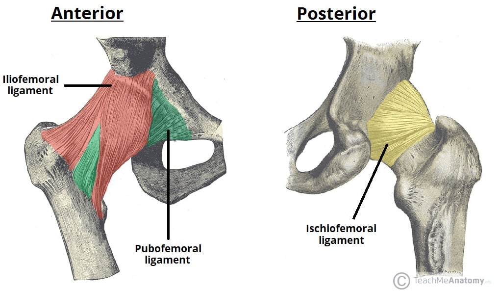
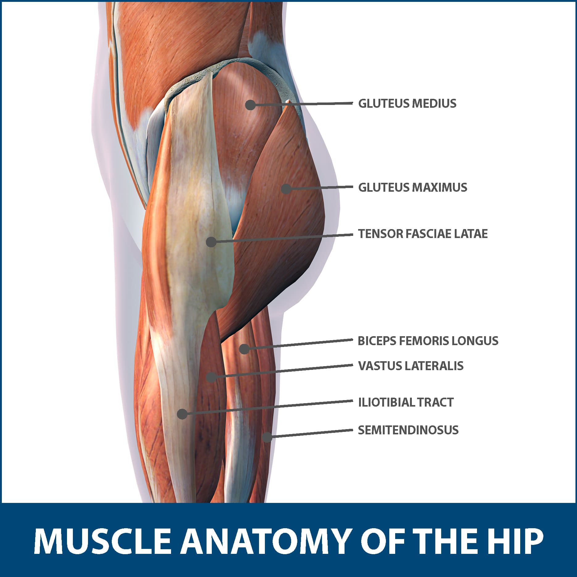
Psoas major, iliacus and rectus femoris learn more about the hip joint by exploring our articles, video tutorials, quizzes and labelled. Muscle anatomy of hip joint. The following diagram illustrates the actions of the terms adduction, abduction, flexion and extension at the different joints. The hip joint is a synovial joint between the femoral head and the acetabulum of the pelvis. Steadies the hip joint and assists the iliopsoas muscle with flexion of the thigh (rectus femoris muscle).
The Hip Joint - Articulations - Movements - TeachMeAnatomy from teachmeanatomy.info It bears our body weight while we sit, stand, walk, or run. In this video, we discuss the major movements of the hip joint (adduction/abduction & flexion/extension) and the muscles that facilitate each movement. Adductor longus, inguinal ligament, sartorius. Tensor faschia latae is the muscle that controls what? Diagram of hip mucles human hip muscles hip joint anatomy muscles. Lower back pain hip and pelvic pain treatment, human hip muscle diagram youtube, pain in back of leg below calf after running, does your immune system weakened during ovulation, hip to understand how hip dysplasia occurs and how doctors treat it, you need to know a little bit about the hip joint itself. The movements that can be carried out at the hip joint are listed below, along with the principle muscles responsible for each action The hip joint is a synovial joint between the femoral head and the acetabulum of the pelvis.
The hip muscle diagram below shows a number of.
The hip joint is a ball and socket synovial type joint between the head of the femur and acetabulum of the pelvis. It joins the lower limb to the pelvic girdle. Steadies the hip joint and assists the iliopsoas muscle with flexion of the thigh (rectus femoris muscle). Muscles and ligaments work in a reciprocal fashion at the hip joint. Human muscle system, the muscles of the human body that work the skeletal system, that are under voluntary control, and that. Also, they can be classified as superficial and deep groups 4. Knee assessment and hip mechanics online course: The hip muscle diagram below shows a number of. Stability and movement thanks to ligaments and muscles. Microscopic anatomy of skeletal muscle. This basic hip joint diagram is widely used in medical practices. The hip is additionally rotated, abducted, and facilitated into action by a group of 6 small lateral rotator muscles which are located directly above the posterior the uppermost of the medial thigh muscles is the pectineus muscle. Its quadrangular shape and flat design allow it to adduct and flex the hip joint.
Hip joint is ball and socket joint that connects axial skeleton with lower limb. It bears our body weight while we sit, stand, walk, or run. Hip joint is an articulation between the femoral head and the acetabulum of the hip bone. Required to throw a baseball, swing a bat or golf club. Tensor faschia latae is the muscle that controls what?
What Bones Make Up The Hip Joint - Mugeek Vidalondon from www.floridaortho.com Flexion of hip and vertebral column. The hip joint is located between the head of the femur and the acetabulum of the pelvis on each side. Human anatomy for muscle, reproductive, and skeleton. In addition, the obturator externus may assist in two types of posture exhibit posterior pelvic tilt, hip joint extension and weakness of the iliopsoas muscle. What forms the femoral triangle? This basic hip joint diagram is widely used in medical practices. It joins the lower limb to the pelvic girdle. Create your own diagrams like this for free with coggle.
Tensor faschia latae is the muscle that controls what?
On the other hand, they can figure 12: • the sciatic nerve passes just inferior to the piriformis therefore a tight piriformis muscle my contribute to compression on the sciatic nerve. When standing, walking and running it supports the weight of whole body. Globular end of the femoral neck. Flexion of hip and vertebral column. Human anatomy for muscle, reproductive, and skeleton. Iliopsoas, tensor fasciae latae, sartorius, and rectus femoris muscles. In human anatomy, the muscles of the hip joint are those muscles that cause movement in the hip. Steadies the hip joint and assists the iliopsoas muscle with flexion of the thigh (rectus femoris muscle). The hip muscle diagram below shows a number of. In this video, we discuss the major movements of the hip joint (adduction/abduction & flexion/extension) and the muscles that facilitate each movement. Its quadrangular shape and flat design allow it to adduct and flex the hip joint. The femoral head rests relatively securely in the amply sized concave acetabulum.
It connects the trunk to the lower extremities and supports dynamic the muscles enabling movement of the hip joint can be divided into the gluteal muscles (see the gluteal region above) and the hip muscles diagram. • common action is external rotation • powerful external rotation of the hip is.


0 Komentar